Research Theme
9. Detailed distribution of sphingomyelin on the plasma membranes revealed by specific probes
Motohide Murate, and Toshihide Kobayashi
Lipid Biology laboratory, RIKEN
Recently, specific sphingomyelin (SM)-binding toxin, equinatoxin II (EqtII) was developed as a probe. Biochemical analysis using model membranes with different lipid composition showed this protein preferentially binds to dispersed SM molecules. It has been well characterized that another SM-binding probe, lysenin, binds to SM clusters of 5-6 molecules [1]. Detailed distribution of sphingomyelin on the plasma membrane of various cells was analyzed using both probes in immunoelectron microscopy.
Madin-Darby canine kidney (MDCK) cells polarized into apical and basolateral membranes when they were cultured in high density. Each plasma membrane has a specialized function and contains a different set of lipids and proteins. So, we compared the distribution patterns of lysenin and EqtII in plasma membrane of differentiated and undifferentiated cells. Lysenin bound to undifferentiated membranes of MDCK cells in low density, whereas labeling was almost not observed in apical membranes of differentiated cells. On the contrary, weak binding of EqtII in undifferentiated membranes was dramatically increased in differentiated apical membranes (Fig. 1). These results indicate that a SM distribution pattern in plasma membrane change after the differentiation of cells as epithelium.
Niemann-Pick disease type A (NPA) is a genetic defect of acid sphingomyelinase characterized by the accumulation of SM in late endosomes/lysosomes. The cell surface distribution of the lipid in NPA cells is not well understood. Thus, the labeling of normal and NPA fibroblasts to lysenin and EqtII was investigated. As show in Fig. 2, lysenin labeling in the plasma membrane of NPA cells was dramatically decreased, whereas EqtII equally labeled in normal and NPA cells. Biochemical analysis also demonstrated that cell surface sphingomyelin content was significantly lower in NPA fibroblasts compared to normal skin fibroblasts [2].
 Fig. 1. Cell surface labeling of Lys and EqtII-GFP were estimated by
calculating the density of gold particles on the plasma membrane
sheets from MDCK cells in low and high cell densities.
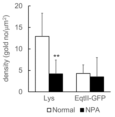 Fig. 2. Cell surface labeling of Lys and EqtII-GFP were estimated by calculating the density
of gold particles on the plasma membrane sheets from normal and NPA fibroblasts.
【References】
[1] Ishitsuka, R. et al. (2004) Biophys. J. 86, 296–307.
[2] Makino A., Abe M., Murate M., et al. (2015) FASEB. J. 29, 477–493.